Patellar Instability and Common Risk Factors
Patellar instability is a condition characterized by the abnormal movement or dislocation of the patella (kneecap) from its normal position.
It accounts for about 3% of all knee injuries.
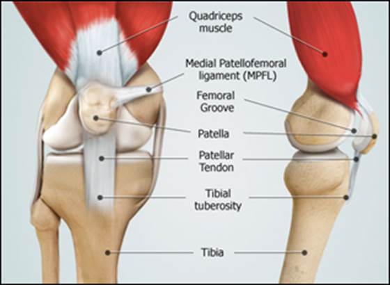
The patella is attached to the femur (thigh bone) and tibia (shin bone) by the quadriceps and patellar tendons. It sits in a groove at the end of the femur known as the trochlear groove where it slides up and down as the knee bends and straightens.

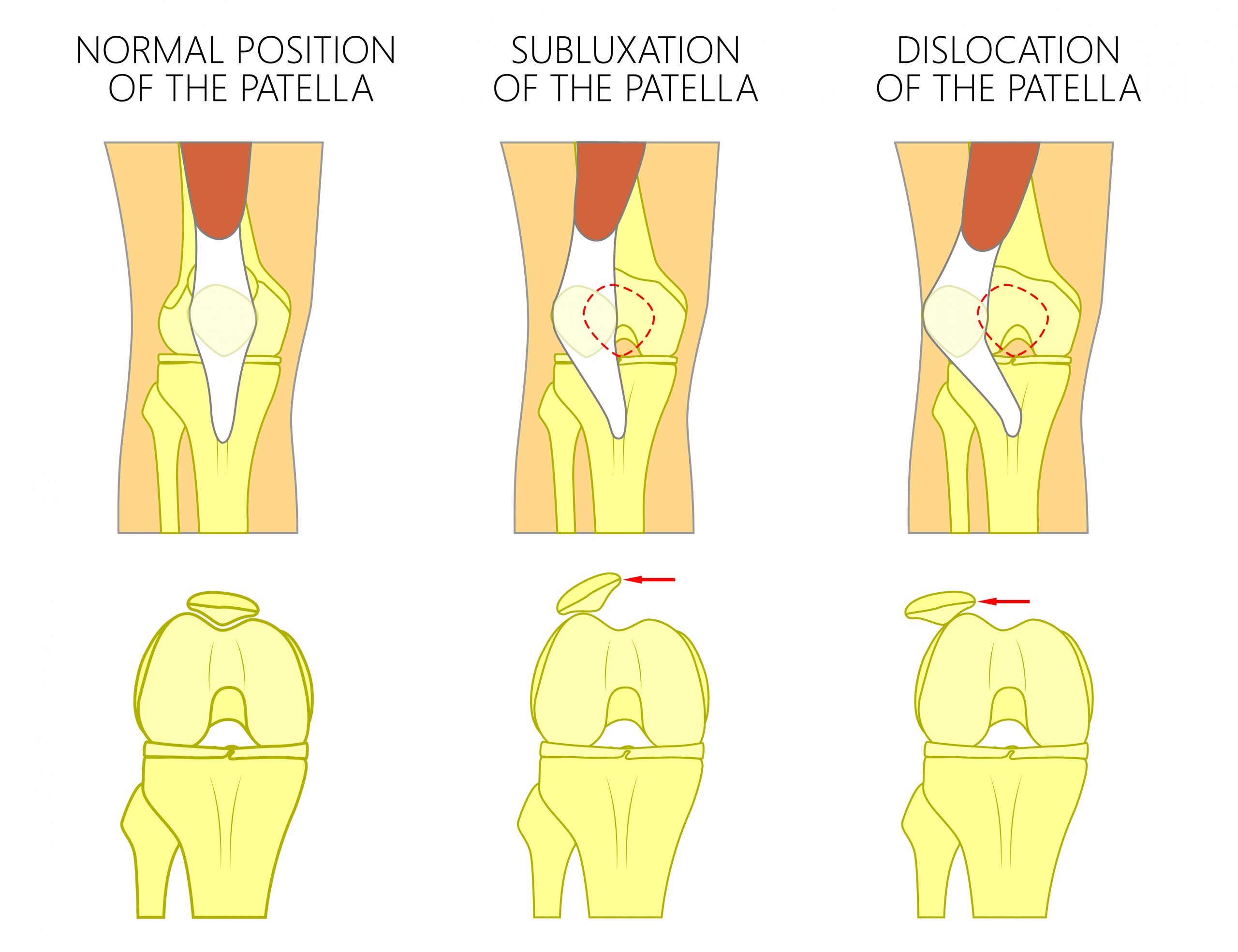
When the patellar displaces, it can slide partially out of the groove, called subluxation, or it can get pushed completely out of the groove and dislocate.
Displacement of the patellar, whether it subluxes or dislocates, can result in pain, swelling, instability, and difficulty with knee movements.
— Learn more about the basics of patellar instability —
Common risk factors for patellar instability
While patellar instability can occur in anyone, it is more common in young females.
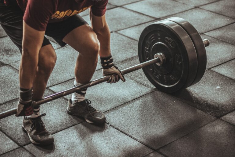
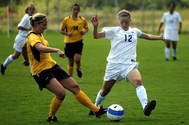
It can occur due to injury, such as a direct impact to the knee from playing high impact sports or activities, or from variations in a person’s anatomy, such as a shallow or uneven trochlear groove, loose ligaments or extremely flexible joints, that allow the patella to displace with minimal force.
Below are some of the risk factors that predispose some individuals to patellar instability more than others.
Patella Alta
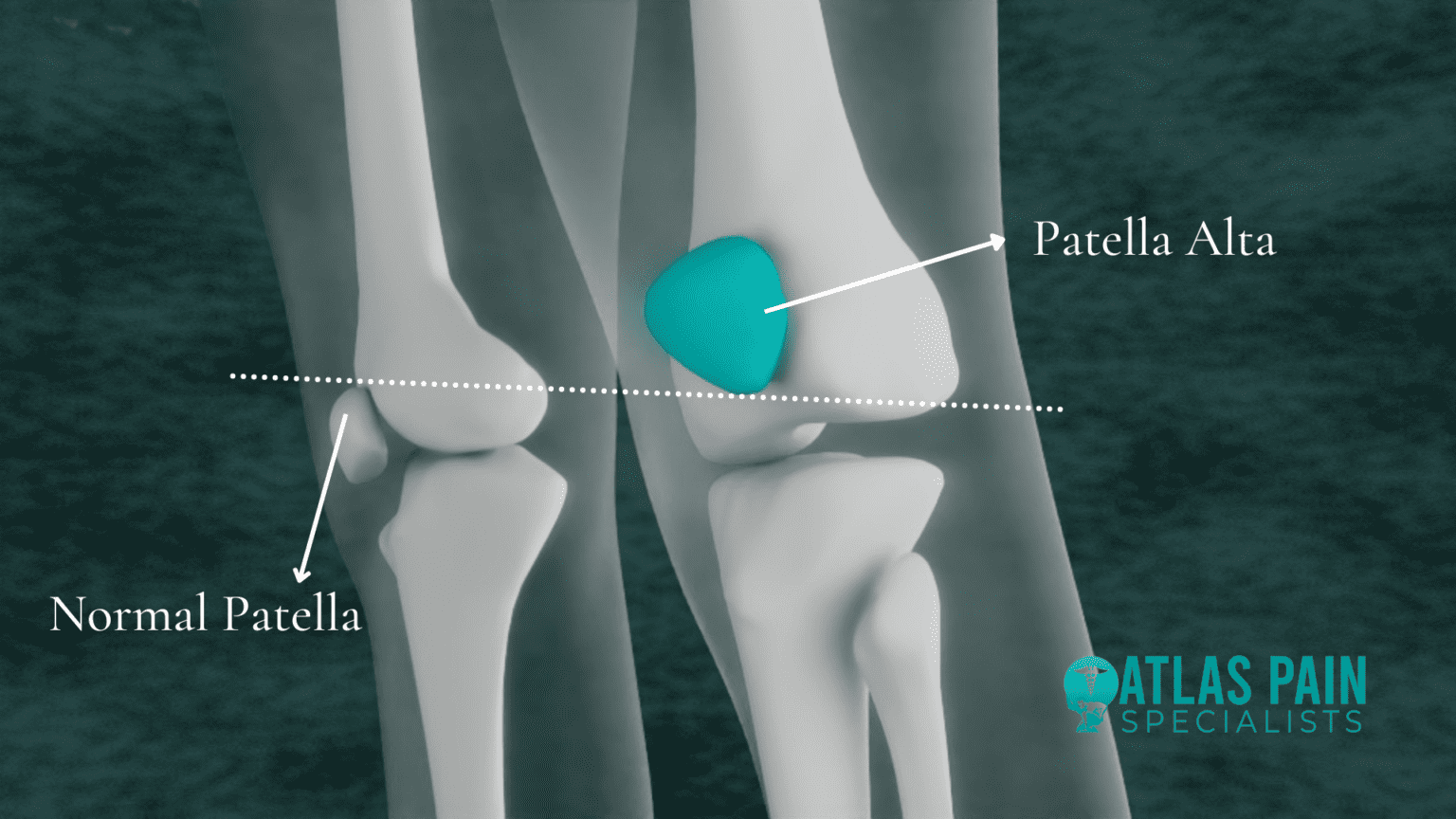
Patella Alta (high-riding patella) refers to a higher than normal position of the patella in relation to the femur where the trochlear groove is very shallow.
Because of this, the groove provides only a small barrier on each side to keep the patella in place. Hence, the patella is at high risk of being pulled sideways over the low edge of the groove as the knee bends and can partially or fully dislocate out of position.
While patella alta can result from various causes including injury, for the most part, this condition is caused by congenital/developmental defects where the patellar tendon in naturally longer than average.
Trochlear Dysplasia
Trochlear dysplasia refers to an abnormal, relatively flat or dome shape of the femoral trochlea, where the patella loses stability and can track to the outside of the knee when the knee bends.
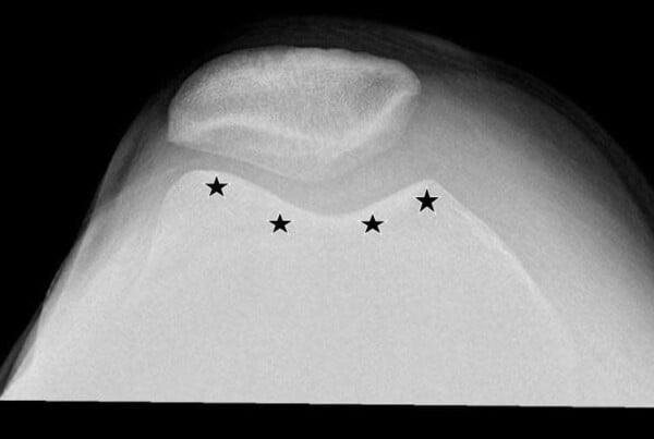
Image: https://drrobertlaprademd.com/
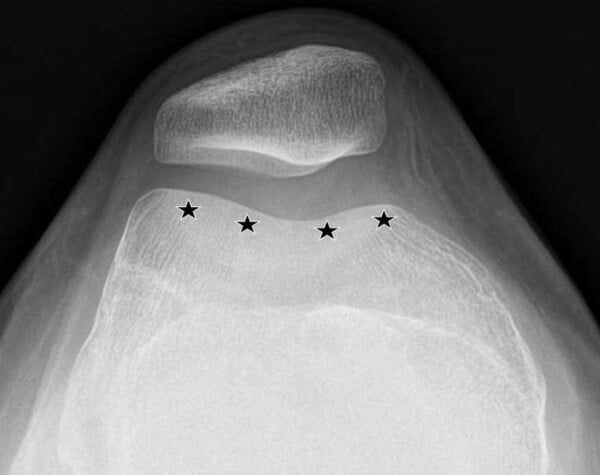
Most cases of trochlear dysplasia are believed to be genetic. However, there have been cases in younger individuals where knee dislocations that aren’t put back into place, later affect the proper development of the trochlear groove.
Studies have shown a strong association between trochlear dysplasia and patellar instability, as those who have some form of trochlear dysplasia have a much greater rate of recurrent dislocations, depending upon how flat their trochlear groove is.
Medial Patellofemoral Ligament Deficiency
The medial patellofemoral ligament (MPFL) is the main stabilizing ligament of the patella that prevents the patella from dislocating to the outside of the knee.
Deficiency of the MPFL can occur either through injury, where the MPFL gets damaged or torn, or in people with ligamentous laxity (loose ligaments). In these instances, the MPFL is not able to effectively hold the patella in place to stop it displacing sideways.
Ligaments are the tough fibrous connective tissue that holds your bones together at joints and keeps the joint stable.
— Learn about MPFL repair/reconstruction —
Previous Patellar Dislocation
A previous patellar dislocation is one of the most significant risk factors for recurrent instability, especially in patients with an MPFL injury. Once the patella has dislocated, it can disrupt the anatomical structures causing pain, functional limitations, instability, and increase the likelihood of future dislocations.
Contributing Expert

Maria Mohorea, Third Year Health Science Honors Student, McMaster University
References
Dejour, H., Walch, G., Nove-Josserand, L., & Guier, C. (1994). Factors of patellar instability: an anatomic radiographic study. Knee surgery, sports traumatology, arthroscopy : official journal of the ESSKA, 2(1), 19–26. https://doi.org/10.1007/BF01552649
Hayder Adnan, S., Marwan Jwad, M., & Rafal Hani, M. (2021). MRI evaluation of trochlear dysplasia as a cause of patellar instability. International Journal of Radiology and Imaging Techniques, 7(2). https://doi.org/10.23937/2572-3235.1510087
Kelly, B. T., Maak, T. G., Larson, C. M., Bedi, A., & Zaltz, I. (2013). Sports hip injuries: assessment and management. Instructional course lectures, 62, 515–531.
Patella alta – causes & treatment. Knee Pain Explained. (2022). https://www.knee-pain- explained.com/patella-alta.html
Patellofemoral pain syndrome (PFPS) treatment in NJ: Pain management doctor. NJ’s Top Orthopedic Spine & Pain Management Center. (2023). https://redefinehealthcare.com/patellofemoral-pain-syndrome/
Wolfe, S., Varacallo, M., Thomas, J. D., Carroll, J. J., & Kahwaji, C. I. (2023). Patellar Instability. In StatPearls. StatPearls Publishing.