Why is my hip sore Doc?
Banff Sport Medicine Physician, Dr Andy Reed, addresses this question in his latest Bow Valley Crag & Canyon article.
Read the original article here
I see a lot of painful hips in my clinics.
One of the commonest problems I see is gluteal tendinopathy, although I find most of my patients know this as ‘Hip Bursitis’.
The typical patient is a female in the 50s or older. There is usually no history of a specific injury though sometimes there has been a fall onto the outside of the hip.
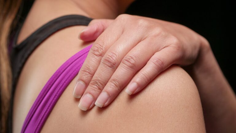
The patient has usually had pain for several weeks or months, located on the outside of the hip, with a tender area at the bony prominence of the hip (the greater trochanter) or just behind it. There may be pain that radiates down the outside of the hip towards the knee, but usually the sore spot is on the outside of the hip.
Typically patients report that lying on the affected side at night causes the most pain, but even lying on the opposite side can be painful, especially if the upper leg adducts across the lower leg, compressing the structures on the outside of the hip.
There is often pain getting up from sitting, especially from lower chairs, and getting in and out of the car can be difficult. Patients often report that pain improves as they get going, but will worsen if they do too much, and often the pain is bad after prolonged activity.
Bursitis or tendinopathy?
A quick google search, which most of my patients have performed, will reveal a possible diagnosis of ‘hip bursitis’.

A bursa is a small lubricating sac that exists between tendons, or between tendons and bone, or bony prominences. We have lots of different bursae in the body, and several commonly exist on the outside of the hip.
Bursae are full of nerve endings, and any inflammation in these structures is usually painful.

Most people with lateral hip pain, however, do not have an inflamed bursa, and we know this from our clinic ultrasound scans or MRI studies. Typically the pathology is a gluteal ‘tendinopathy’.
What is gluteal tendinopathy?
Tendinopathy refers to tendon damage and can encompass anything from an acute overuse injury (tendinitis) to a tendon rupture.
Fortunately in most cases of gluteal tendinopathy, there is no large rupture, but often micro-tears of the either the gluteus minimus or gluteus medius tendons where they insert onto the outside of the femur at the greater trochanter.
Sometimes the bursa between these tendons becomes inflamed, hence ‘bursitis’ but more often than not the bursa is normal, and all we see on ultrasound is damage to the tendon, with small areas of damage or calcifications.
What causes gluteal tendinopathy?
We don’t fully understand why damage occurs at tendon insertions onto bone, but as we age, cumulative trauma, overuse and potentially other conditions such as vascular disease can affect the health of our tendons.
After menopause, women are much more likely to develop tendon injuries, and this together with different hip biomechanics makes older females most susceptible.
Injuries are far more likely to occur in weakened tendons and their associated muscles, so strength training is always part of the treatment.
Diagnosis and treatment
In clinic we will examine you, usually perform an ultrasound of the hip area, and order xrays, as hip arthritis can present with lateral hip pain.
Once we diagnose gluteal tendinopathy or hip bursitis, exercises will be typically prescribed. These may include simple home exercises such as gluteal bridges, clam shells and resistance band exercises to strengthen the hip abductors. Often a physiotherapist will supervise the progression.

Sleeping with a pillow between the knees can provide relief at night, and typically we advise limiting any obviously aggravating sports.
In persistent cases we may recommend injections, either steroid injected into the bursa using ultrasound to guide needle placement, or a newer treatment like platelet rich plasma directly into the injured tendon; usually these are second-line treatments that we only consider after a few months of strengthening.
A less invasive treatment called shockwave therapy is also gaining popularity, and is often available through physiotherapy offices. Powerful shockwaves are directed at the damaged tendon, and are believed to stimulate tissue regeneration through a number of mechanisms. It’s a mildly uncomfortable treatment that is repeat over several weeks and can be very effective.
Regardless of any additional treatments, we will always recommend strengthening.
Lastly, if all conservative options have been exhausted, MRI imaging can be useful to determine if surgery could be helpful, but in my experience it is very unusual to need surgery.
Contributing Expert
Dr Andy Reed, Banff Sport Medicine Physician